What is femur and femoral neck
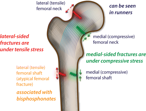
Hip is a ball and socket joint where upper leg meets the pelvis. Femur is the longest, strongest bone of the body and is also called “thighbone”. At the top of the femur is the femoral head. This is the ball that sits in the socket. Just below the femoral head is the femoral neck. Because femur is a weight bearing structure and femoral neck is the narrow part, stress fractures occur in this area. The importance of femoral neck stress fractures is that if detected early, and the injury is un-displaced, management and outcome have much higher success rates, when compared to displaced fractures.
HOW IT OCCURS
When bone is subjected to repetitive daily subthreshold loading, micro-cracks may occur within cement lines. The normal re-modelling a process repairs these cracks.
However, if the bone continues to be subjected to high stresses, then crack propagation occurs. If crack progression is faster than the repair, it will become a stress fracture. Stress fractures occur when normal bone is exposed to abnormal stress.


RISK FACTORS
They are seen in professional athletes and in military personnel.
Activities like long distance running, abrupt increase in training without adequate rest may predispose to stress fractures.
Women are at increased risk of stress fractures because of narrower bones and lower bone mineral density, especially in postmenopausal women with osteoporosis.
Calcium deficiency and vitamin D deficiency.
Smokers are prone for more these fractures
NSAIDs use, overweight, muscle weakness or imbalance predispose to stress fractures.
PRESENTATION
Most common symptom is gradual onset hip or groin pain that is aggravated by activity and weight bearing and ceases with rest.
Patients often notice a recent increase in intensity or duration of exercise, such as preparation for a marathon or a sports event. Initially, pain is noted late in an activity, eventually pain is noted at rest and at night.
On examination pain on extremes of hip range of motion, especially with internal rotation. Tenderness over front of thigh and the inguinal area. Pain can be increased by doing straight leg raise test.
DIAGNOSIS
X-RAY can be useful if a stress fractures are seen, then further imaging is rarely necessary.
However, x-ray may be false negative for up to 3 months after symptoms onset


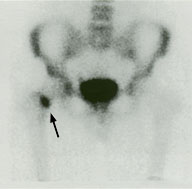
Scintigrams (isolated bone scans) are very sensitive, but not specific.


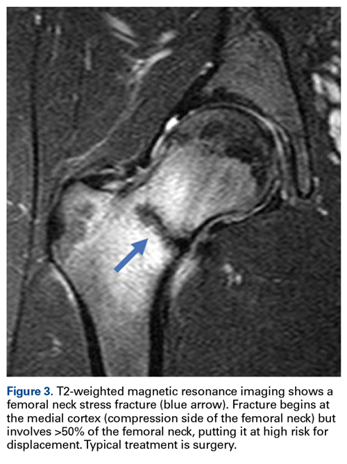
MRI is both specific and sensitive for a stress fracture and involves no radiation exposure.


TREATMENT
There are nonoperative and operative treatment plans are available.
Nonoperative treatment includes non weight bearing, crutches and activity restriction. Majority of stress fractures can be successfully treated non-operatively by avoiding stress activity.
Operative techniques include percutaneous screw fixation. Operative techniques are used for cases of delayed union or failed non-operative treatment.
Frequently Asked Questions
